Each Community Spotlight features an outstanding group, partner, resource, or member of our community.

Bioimage Informatics Activity

By David Julian

Cell culture images


Module Description:

In this two-part module, students use basic bioimage informatics techniques to acquire quantitative data from images of cultured cells, and then use these data to test a hypothesis about the effect of a genetic mutation on cellular phenotypes. Together, the two activities are designed to model an authentic research project that uses modern bioimage informatics techniques.

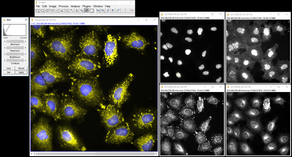
In the first part of the module, students analyze fluorescence emission images of cultured cells to determine the appearance, characteristics, and dimensions of some cellular features, and then create a composite, color image. In the second part of the module, students use image analysis to count and measure the nuclei of control cells and cells with a mutation in a proto-oncogene, and then use a t-test to test for statistical significance.


Teaching Setting:

The module is intended for students in a college-level general biology course, but is also suitable for a cell biology course.  Each part takes about two hours and is intended to be performed in a computer lab (or any classroom or laboratory in which students have access to computers), but it may be feasible for students to perform the exercise at home using a personal computer. The required software is freely and publicly available, and can be installed and run on a computer running Windows, Mac OS, or Linux, or can be launched directly in a browser from the ImageJ Software page on QUBES.  Students may work individually or in pairs.


Citation:

Julian, D. (2018). Bioimage Informatics Activity. HHMI BioInteractive FMN (2016), QUBES Educational Resources. doi:10.25334/Q49M6M


Visit Resource


Share
Tweet


Related Materials and Opportunities:

The QUBES Open Education Resources (OER) Collection now includes more than 800 resources!  QUBES has an OER sharing platform that makes it easy to share your work and track its impact.  The QUBES Resources System assigns a stable Digital Object Identifier (DOI) to each resource making it easy to track and find your resource, and measures the use of each resource including views, downloads and adaptations.  For example, the resource featured in this ROW is an adaptation of the HHMI BioInteractive Cancer Discovery Activities (available on both the HHMI BioInteractive site and on QUBES) and at the time that this ROW was published, this resource had more that 240 views and 150 downloads!  Learn more about OER on QUBES.

In this resource, students process images of cultured cells using the image processing software ImageJ, which is one of several software hosted by QUBES.  There is no need to have students download the software - they can launch it directly in their browser from the ImageJ Software page on QUBES, saving precious class time.  This resource is also currently under development to make it even easier for students to access the images used in this activity through ImageJ.  Be sure to “watch” this resource so you’ll be notified when a new version is available!  

The author developed this resource while participating in the 2016 HHMI BioInteractive Faculty Mentoring Network (FMN).  This resource is one of many resources that have been customized or extended for use in a variety of classroom settings.  Browse all HHMI BioInteractive Educator Publications.   

In addition to the adaptations described above, several original HHMI BioInteractive resources have been added to the QUBES Collection of open education resources in just the past few weeks.  They are listed below.  Check them out! 

  • “Cells of the Immune System”, a tutorial that provides an overview of the immune system, concentrating on the roles played by B and T lymphocytes, and on the antigen-presentation system.

  • “Targeting Infected Cells for Immune Defense”, an animation that shows how a cell infected by a virus signals cytotoxic T cells to destroy itself.

  • “The p53 Gene and Cancer”, a tutorial that describes the structure and function of the p53 protein, how its activity is regulated in cells, and how mutant versions of p53 can lead to cancer.

  • “Cystic Fibrosis Mechanism and Treatment”, an animation that shows how mutations in an ion channel protein lead to the genetic disease cystic fibrosis. The animation also discusses how research on this protein has been used to develop treatments for the disease.

  • “Sex Verification Testing of Athletes”, an interactive module that explores the biology of sex determination and development in humans, set against the backdrop of the different sex testing policies implemented throughout sports history.

  • “The Biology of Skin Color”, a film that explores the hypothesis that different tones of skin color in humans arose as adaptations to the intensity of ultraviolet radiation in different parts of the world.

  • “Natural Selection in Humans”, a film that explores the evolutionary connection between an infectious disease, malaria, and a genetic condition, sickle cell anemia.

  • “Electrical Activity of Neurons”, a tutorial that describes how neurons generate action potentials, and how scientists measure neuronal activity and record the firing of individual neurons.


QUBES on Social Media
BioQUEST is a transformative, collaborative community empowering educators to drive innovation in STEM education for all students.
Copyright © 2024 QUBES, All rights reserved.
P.O. Box 1452, Raymond, NH 03077
You are receiving this email because you have shown interest in receiving updates from BioQUEST and QUBES.

Subscribe / Unsubscribe from mailing list
View Community Spotlight on QUBESHub
Community Spotlight: Issue 46